
Where and when an artifact was created
Accelerated ion beams for art forensics
Philippe Collon and Michael Wiescher
Source - http://www.physicstoday.org/resource/1/phtoad/v65/i1/p58_s1?bypassSSO=1
The worlds of art and archaeology have adapted techniques developed in nuclear physics laboratories to learn where and when an artwork or artifact was created.
A couple of months ago, in a courthouse in Cologne, Germany, four people were sentenced to a total of 15 years in prison for art forgery. As one art historian exclaimed during the trial, “It is the most exciting scandal in the art world after [World War II].” The four had been on trial for selling about 50 paintings, mostly of German surrealists and expressionists from the early 20th century, from the fictitious Werner Jägers and Wilhelm Knops collections; they admitted to being involved in 14 forgeries. Confessed-to imitations included faked works of such important artists as Heinrich Campendonk, Max Ernst, and Fernand Léger. The total sales of the bogus art have reached about €27 million ($36 million). All the fakes were certified as original by art experts who relied on their knowledge of the artists and their work.
The trial clearly illustrates the folly—particularly in a society in which art can be a major investment—of relying entirely on art historians and others who claim that their expertise makes any scientific investigation pointless. Art experts play an important role in identifying the style, history, and context of a painting, but a solid scientific basis for the proper identification and classification of a piece of art must rely on information from other sources.
For example, investigators working the German forgery case used tree-ring dating to prove that the same tree provided wood for the frames of four paintings supposedly from different artists: Collioure by André Derain, Nature morte by Léger, Else Lasker-Schüler gewidmet by Campendonk, and Seine mit Brücke und Frachtkähnen by Max Pechstein. A second technique, proton-induced x-ray emission (PIXE), enabled investigators to study the composition of paint pigments used in the forgeries.
Accelerators in the art world
A host of approaches with origins in biology, chemistry, and physics have allowed scientists and art historians not only to look below a painting’s or artifact’s surface but also to analyze in detail the pigments used, investigate painting techniques and modifications done by the artist or by art restorers, find trace materials that reveal ages and provenances, and more. Those techniques can provide a slew of information to help substantiate or negate the authenticity of an artwork or artifact, and they also furnish information essential for careful restoration and preservation. In particular, nuclear physics has provided numerous analysis and detection techniques that involve accelerated ion beams from small to midsized accelerators such as electrostatic accelerators or cyclotrons. Key innovations include the capability to deliver well-focused particles and to identify trace elements to a very high degree of sensitivity.
Several accelerator-based laboratories operate solely to make beam techniques available to the worlds of art and archaeology and to others with a need for material analysis. (For industrial applications, see the article by Robert Hamm and Marianne Hamm in PHYSICS TODAY, June 2011, page 46.) Well-known examples are the Accélérateur Grand Louvre d’analyse élémentaire, at the Louvre Museum in Paris; the Laboratorio di Tecniche Nucleari per i Beni Culturali in Florence, Italy; and the Centro Nacional de Aceleradores in Seville, Spain. Most of the facilities are in Europe and are dedicated to investigating and maintaining the continent’s cultural heritage.
It is beyond the scope of this Quick Study to review all the accelerator-based methods presently being used for art analysis. However, two techniques that we employ at the University of Notre Dame should illustrate some of what’s possible.
PIXE, a nondestructive analysis tool
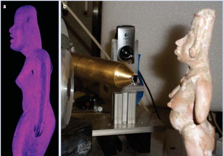
The two related figurines shown here were found in Mesoamerican burial grounds and are currently housed at the University of Notre Dame’s Snite Museum of Art. (a) Exposing an artefact to UV light reveals the location of pigments invisible to the unassisted eye. (b) Proton-induced x-ray emission (PIXE) provides quantitative details of the pigment composition; in particular it reveals the iron and manganese content of the paint. Seen here is the copper-colored PIXE beam line from Notre Dame’s 11-MV electrostatic accelerator. Just visible at the left is the silicon–lithium detector used for observing the characteristic emitted x rays. Techniques such as UV and PIXE analysis are helping anthropologists trace two millennia of Mesoamericancultural and religious history.
When a proton beam interacts with the material of a sample, it can liberate atomic electrons from inner shells. As other electrons relax to fill the vacancy, they emit electromagnetic radiation, mostly x rays. PIXE is a technique that determines the elemental composition of the sample through the detection of that characteristic electromagnetic radiation. In that regard, it is similar to another common technique called x-ray fluorescence. But PIXE has several advantages over x-ray fluorescence. Varying the beam energy enables depth profiling of a sample, and employing thin beams allows for microscopic spot analysis. Moreover, PIXE has a much lower noise-to-signal ratio than does x-ray fluorescence, an advantage that makes it possible to observe a whole range of trace elements not previously identifiable.
The scope of applications of PIXE in the art world has grown steadily. When applied to a work of art, as shown in the figure, PIXE helps to identify the composition of pigments or other materials; thus it has had a growing impact in the forensic analysis of suspected forgeries. The analysis of ancient coins provides information about the minting process and also leads to deeper insight into economic developments. For example, inflation during the Roman Empire is reflected in a continuous devaluation of the silver denarius coin, as silver was gradually replaced by less valuable metals. In collaboration with others at Notre Dame, we are investigating the unique black-and-white ceramics of the American Southwest to identify whether mineral or organic pigments have been used to generate the paint and to determine the provenance and distribution of the pottery material. We have also joined with our colleagues to explore the frequently shifting 18th-century colonial boundaries in the present US Midwest by studying the composition of regional Native American copper jewelry. With PIXE, copper mined locally in the upper lake region can be distinguished from British or French imported copper.
A well-known case of PIXE assisting comparative dating is its analysis of the ink used in Galileo Galilei’s frequently undated writings and notes. That analysis has led to a better understanding of Galileo’s evolving thought processes and has shed new insight on his role in science history. Another example is the sequencing of silverpoint drawings by German Renaissance painter Albrecht Dürer, who periodically traveled to the imperial court to receive his annual stipend. On those trips he used silverpoint pens, purchased at various locations, to draw local scenery or to record moments and ideas he would later use for his paintings. PIXE compositional analysis allowed scholars to figure out the order in which those drawings were produced, which, in turn, led to an improved understanding of Dürer’s artistic development.
Accelerator mass spectrometry
An isotopic analysis of materials via conventional mass spectrometry can reveal a great deal of information about the provenance of a given sample; indeed, mass spectrometry is a routinely used analysis tool. The innovation of accelerator mass spectrometry (AMS), however, has made it possible for researchers to measure trace radionuclides in tiny samples with unprecedented sensitivity.
As with conventional mass spectrometry, AMS identifies a specific radionuclide of interest by separating it from other isotopes with different mass-to-charge ratios. But in AMS, an accelerator provides energy to the ions released from the sample. The high-energy ions are then amenable to standard nuclear physics techniques that detect, for example, time of flight, energy E, stopping power (the rate dE/dx at which energy is lost with distance xas a particle passes through a medium), and position in a focal plane. Such information determines an isotope’s mass number A and atomic number Z and allows for the identification of a specific isotope, even if it is present in an isotopic (same Z) and isobaric (same A) background that is higher by as much as 16 orders of magnitude.
Accelerator mass spectrometry is now the tool of choice for the dating of archaeological artifacts (see the article by Ervan Garrison in PHYSICS TODAY, October 2001, page 32). That’s in part because the method requires such a small amount of material to achieve its exceptional precision. Celebrated examples of objects dated by AMS are the Shroud of Turin and Ötzi the Iceman, a well-preserved mummy discovered in 1991 on the Austro–Italian border. In both cases, samples weighing no more than some tens of milligrams were removed from the artifacts and dated via carbon-14 decay. In fact, the degree of precision provided by 14C AMS dating is now inspiring the development of ever-smaller and more user-friendly 14C AMS systems.
The techniques of AMS and PIXE are just two examples from a wide range of nuclear physics applications that could be exploited with existing technology. Such efforts do more than just provide crucial scientific information. Because they are interdisciplinary, they also serve to improve communication and intellectual exchange between scientists and our nonscientist colleagues. That kind of collaboration allows science departments not only to train a number of undergraduate students who would not traditionally be associated with a nuclear research laboratory and nuclear techniques but also to introduce them to state-of-the-art equipment and demonstrate the societal benefits of nuclear physics.
Philippe Collon is an associate professor of physics and Michael Wiescher is a Frank M. Freimann Professor of Physics at the University of Notre Dame in Notre Dame, Indiana.
References
- G. Amsel, M. Menu, J. Moulin, J. Salomon, “The 2 MV tandem pelletron accelerator of the Louvre Museum,” Nucl. Instrum. Methods Phys. Res. B 45, 296 (1990). [Inspec]
- P. Mandó, “INFN-LABEC—Nuclear techniques for cultural heritage and environmental applications,” Nucl. Phys. News 19 (1), 5 (2009).
- J. G. Camacho, R. G. Tenorio, “Centro Nacional de Aceleradores (CAN), Sevilla, Spain,” Nucl. Phys. News21 (3), 5 (2011).
- E. Ciliberto, G. Spoto, eds., Modern Analytical Methods in Art and Archaeology Wiley, New York (2000).
- P. Collon, D. Robertson, in The Encyclopedia of Mass Spectrometry vol. 5, D. Beauchemin, D. E. Matthews, eds., Elsevier, Boston (2010), p. 621.
- National Academy of Sciences, Scientific Examination of Art: Modern Techniques in Conservation and Analysis National Academies Press, Washington, DC (2005), online at http://books.nap.edu/openbook.php?record_id=11413.