ARCHEOLOG-HOME / INDIana-UNIversitas
Contact


How is sagittal balance acquired during bipedal gait acquisition?
Comparison of neonatal and adult pelves in three dimensions. Evolutionary implications
Christine Tardieu, Noémie Bonneau, Jérôme Hecquet, Christophe Boulay, Catherine Marty, Jean Legaye, Geneviève Duval-Beaupère
Source - http://www.sciencedirect.com/science/article/pii/S0047248413001346
Journal of Human Evolution
Abstract
We compare adult and intact neonatal pelves, using a pelvic sagittal variable, the angle of sacral incidence, which presents significant correlations with vertebral curvature in adults and plays an important role in sagittal balance of the trunk on the lower limbs. Since the lumbar curvature develops in the child in association with gait acquisition, we expect a change in this angle during growth which could contribute to the acquisition of sagittal balance. To understand the mechanisms underlying the sagittal balance in the evolution of human bipedalism, we also measure the angle of incidence of hominid fossils.
Fourty-seven landmarks were digitized on 50 adult and 19 intact neonatal pelves. We used a three-dimensional model of the pelvis (DE-VISU program) which calculates the angle of sacral incidence and related functional variables. Cross-sectional data from newborns and adults show that the angle of sacral incidence increases and becomes negatively correlated with the sacro-acetabular distance. During ontogeny the sacrum becomes curved, tends to sink down between the iliac blades as a wedge and moves backward in the sagittal plane relative to the acetabula, thus contributing to the backwards displacement of the center of gravity of the trunk. A chain of correlations links the degree of the sacral slope and of the angle of incidence, which is tightly linked with the lumbar lordosis. We sketch a model showing the coordinated changes occurring in the pelvis and vertebral column during the acquisition of bipedalism in infancy. In the australopithecine pelves, Sts 14 and AL 288-1, and in the Homo erectus Gona pelvis the angle of sacral incidence reaches the mean values of humans. Discussing the incomplete pelves of Ardipithecus ramidus,Australopithecus sediba and the Nariokotome Boy, we suggest how the functional linkage between pelvis and spine, observed in humans, could have emerged during hominid evolution.
Figure 1. Sagittal view of a right hip bone and of the vertebral column. The sacral plate represents the boundary between lumbar and sacral vertebrae, its degree of inclination is called sacral slope. The weight (W) of the unit head-arm-trunk is applied to the center of the sacral plate in the adult standing position. The projection of the center of the sacral base is posterior to the center of the hip joint, located at a distance (d). (h): vertical distance from the center of the sacral base to the center of the hip joints. The sacroiliac joint is visible with its center of rotation (black cross). The red dotted lines describe the angle of sacral incidence. Note that we here consider the pelvic frame of reference only. The gray dashed line represents the line of gravity of the trunk in the body frame of reference.
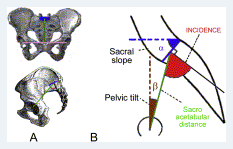
Figure 2. The angle of sacral incidence (A) Frontal and sagittal view of the pelvis. Sacro-acetabular distance is green, biacetabular distance is pink, sacral slope is blue. (B) Angle of sacral incidence, defined by the sacro-acetabular distance -linking the center of the sacral plate and the middle of the biacetabular distance - and the perpendicular to the center of the sacral plate. This angle is the geometric sum of the two positional variables, sacral slope and pelvic tilt. This angle depends upon the degree of the sacral slope, but also measures the degree of the backwards and downwards position of the sacral plate in relation to the acetabula. The variation of the angle of incidence is great.