ARCHEOLOG-HOME / INDIana-UNIversitas
Contact


Eten (Perou): 'Evil Twin' Ovarian Tumor Found In Skeleton From 16th Century
Kristina Killgrove
In an historic cemetery of the Chapel of the Divino Niño Serranito de Eten on the north coast of Peru, bioarchaeologists have discovered nearly 500 burials dating to the Colonial Period. One skeleton in particular, that of a teenage girl, stood out because of the dozens of extra bones and teeth found in her abdominal cavity. Her diagnosis of ovarian teratoma puts her as only the third known example of the phenomenon in ancient history.
Bioarchaeologist Haagen Klaus of George Mason University directs the Lambayeque Valley Biohistory Project, which excavated the ruins of historic Eten between 2009 and 2011. Prior to colonization, Eten was a small community of fishers near the Reque River. Around 1560, local people were rounded up into a colonial settlement called a reducción established to convert the natives to Christianity. Although Eten was abandoned in the mid-18th century, Klaus and his team discovered two churches and their cemeteries underneath sand dunes.
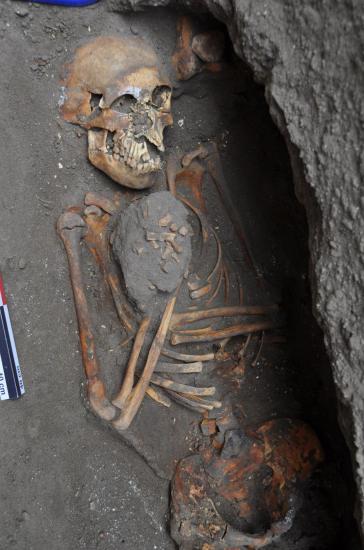 Burial of CNS U2-60, from Colonial-era Eten, Peru, during excavation. This teenager was buried in keeping with the Catholic rite of the time: hands placed on her chest in a praying position. (Photo by Sam Scholes used with kind permission of Haagen Klaus.)
The burial of CNS U2-60 did not at first seem extraordinary, except for its date as one of the earliest burials in Eten. The shape of the skull and pelvis suggest the person was female, and the state of fusion of her long bones means she was in her late teens when she died. She was buried in keeping with the Catholic rite of the time: the body of this young woman was laid out flat, facing the church altar, with her hands placed on her chest in a praying position. As the burial was excavated, though, 83 strange bits of bone and 37 oddly shaped teeth were discovered and were painstakingly documented.
Initially, the mass was thought to be a cultural offering or a grave good, such as a sacrificed animal. But the location of bones in the abdomen and the process of decomposition mean these elements had to be present in this young woman’s body at the time of her death.
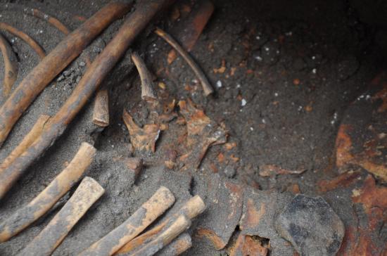 Close-up of the left abdomen of burial CNS U2-60 from Colonial-era Eten, Peru. Remains of an ovarian teratoma can be seen as small flecks between the ribcage and the pelvis. (Photo by Sam Scholes used with kind permission of Haagen Klaus.)
Writing in the International Journal of Paleopathology, Klaus and Connie Ericksen, an archaeology graduate student at Brigham Young University, interpret the anomalous skeletal elements as the remains of an ovarian teratoma. A modern medical term coined from the ancient Greek words for “monstrous tumor,” a teratoma usually presents as a cyst composed of multiple types of cells. This essentially means that a teratoma can grow into a variety of different tissues, including bone, teeth, hair, and organs. While any soft tissue that existed in this ancient case decayed long ago, Klaus and Ericksen were left with a jumble of small fragments of skeletal and dental tissue.
Looking closely at the dozens of extra bony bits, Klaus and Ericksen write that “the general morphology of the bones could be described as unclassifiable in terms of normal human or general mammalian anatomy.” The dental tissue was “approximately the size of human deciduous teeth,” and looked like either small anterior teeth or large molar-like teeth, but “in most cases, occlusal anatomy was an irregular, highly variable arrangement of cusps and cusplets,” they write.
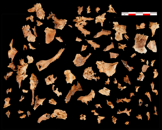 Remains from the ovarian teratoma of burial CNS U2-60 from Colonial-era Eten, Peru, showing skeletal-like tissue. (Photo by Sam Scholes used with kind permission of Haagen Klaus.)
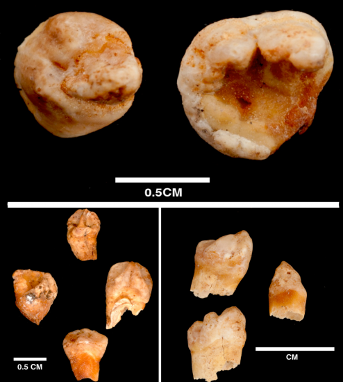 Some of the tooth-like remains of the ovarian teratoma from burial CNS U2-60 from Colonial-era Eten, Peru. (Photo by Sam Scholes used with kind permission of Haagen Klaus.)
The researchers ruled out other diagnoses in this case. The bony remains did not look much like a fetus and there were far too many teeth, so a late-stage ectopic pregnancy is not a good explanation. This means that lithopedion (literally: “stone baby” in Greek), where a dead fetal mass calcifies within the abdomen, can also be ruled out. Fetus in fetu, where a tumor divides into identifiable but parasitic fetal elements, is also unlikely because in this case, there were no defined vertebrae or limbs.
“We know that this type of tumor is fairly common in modern populations, but we have so few examples from prehistory,” bioarchaeologist Allison Foley, who has worked on diagnostic criteria for identifying teratomas in archaeological remains, tells me. They are thought to be present at birth, and most are benign; that is, until they begin to interfere with the body’s functioning. In the case of CNS U2-60, Klaus and Ericksen note that “it is not possible to determine if the lesion was benign or malignant, but a teratoma of this complexity and size likely impacted morbidity via impeded circulation.” She may not have died from the teratoma directly, but the large tumor probably made her appear pregnant and may have factored into her early death.
In spite of the reasonably common finding of ovarian teratomas in modern medicine, ancient evidence of them is practically non-existent. Klaus and Erickson’s finding joins just two previous reports, both from Europe: one from late Roman Spain, and one from Imperial-era Rome. One reason teratomas might not be plentiful in the bioarchaeological literature is that their remains can be easy to misidentify, since they look so much like bone and teeth and can be mistaken for fetal remains. “This case is pretty unambiguous,” Foley says, “thanks to the cluster of 37 teeth found in the abdominal cavity. Even beyond their unusual placement, the teratoma teeth are twisted and malformed.”
With this publication, Klaus and Ericksen want to call further attention to the possibility that this condition was present in skeletal remains of the past, and not just in women. Although this kind of mature teratoma with bone and dental tissues is more commonly found in women, men can also produce testicular teratomas, so all burials need to be excavated with care and with an eye to identifying potential anomalies important for our understanding of health in the past.